Vascular care
10th Jan 2024
Understanding PET Scans for Heart Health
PET Scans for Heart Health
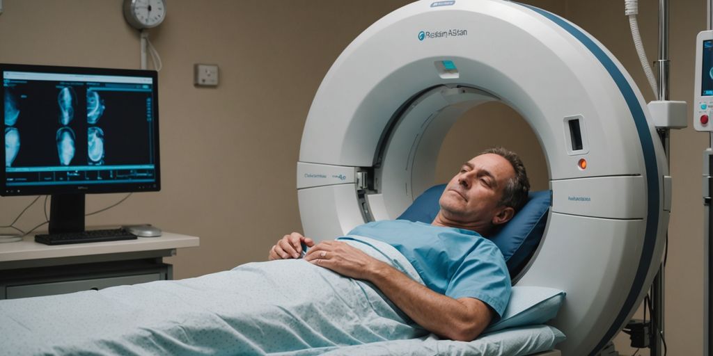
A PET scan for the heart is a special test that helps doctors see how well your heart is working. This test uses a small amount of radiation to take pictures of your heart. These images help doctors understand if your heart is getting enough blood and if there are any damaged areas. This article will help you understand what a heart PET scan is, why you might need one, how to get ready for it, and what to expect during and after the test.
Key Takeaways
- A cardiology pet scan uses a small amount of radiation to take detailed pictures of your heart.
- Doctors use heart PET scans to check for problems like blocked arteries or damaged heart muscle.
- You might need to follow special instructions before the scan, like not eating for a few hours.
- The scan itself is usually done in a hospital and takes about 1 to 3 hours.
- After the scan, you should drink lots of water to help flush out the radiation from your body.
The Basics of PET Scans for Heart Health

What is a PET Scan?
A PET scan of the heart is a noninvasive test that uses radioactive tracers to create images of your heart. This test can define those patients at high risk and who need to undergo procedures like coronary artery bypass surgery (CABG). PET scans help doctors see both healthy and damaged heart muscle, making it a valuable tool in health diagnostics.
How PET Scans Work
During a PET scan, a small amount of radioactive tracer is injected into your bloodstream. This tracer releases energy in the form of gamma rays, which are detected by a special camera. The camera takes pictures of your heart from different angles, creating detailed images. These images help doctors understand your heart’s blood flow and overall cardiac function.
Differences Between PET and Other Imaging Tests
PET scans are unique because they show how well your heart is working, not just its structure. Unlike other tests, PET scans can measure myocardial perfusion and ventricular function. This makes them especially useful for diagnosing conditions that other imaging tests might miss.
When is a PET Scan Recommended for Heart Issues?
Common Symptoms Leading to a PET Scan
Doctors may suggest a PET scan if you’re showing signs of heart trouble. These symptoms can include:
- Irregular heartbeat (arrhythmia)
- Chest pain or tightness
- Difficulty breathing
- Weakness or fatigue
- Heavy sweating
Conditions Diagnosed by PET Scans
A PET scan can help diagnose several heart conditions, such as:
- Coronary artery disease (CAD): This is when the arteries that supply blood to your heart become hard or narrow.
- Heart attack: Cardiology PET scans can show how much damage the heart has suffered.
- Heart infections and diseases like cardiac sarcoidosis.
- Evaluating the health of the heart for a possible transplant.
Comparing PET Scans to Other Diagnostic Tests
PET scans can offer faster, clearer images for diagnosing heart issues compared to other tests. They are often used when other tests like echocardiograms or stress tests don’t give enough information. PET scans are particularly useful in cardiology for mapping blood flow and spotting areas where blood isn’t flowing well.
It’s important for cardiologists to have a clear image of the heart to determine the extent of the plaque buildup or whether the plaque is causing any blockage.
In summary, PET scans are a valuable tool in diagnosing and managing heart disease, offering detailed images that other tests might miss.
Preparing for Your Heart PET Scan
Pre-Scan Guidelines
Your doctor will give you specific instructions to follow before your heart PET scan. It’s important to follow these guidelines closely to ensure accurate results. Generally, you should:
- Avoid eating or drinking anything except water for 4-6 hours before the test.
- Stay away from caffeine, alcohol, and strenuous activity for at least 4-6 hours beforehand.
- Inform your doctor about all medications, including over-the-counter drugs and supplements.
Medications and Dietary Restrictions
Certain medications and foods can affect the results of your PET scan. Here are some key points:
- Medications: Bring a list of all medications you take. Some may need to be paused before the test.
- Dietary Restrictions: Avoid caffeine for 24 hours before the test. This includes coffee, tea, soda, and even chocolate.
- Special Diets: If your doctor is checking for specific conditions like cardiac sarcoidosis, you may need to follow a high-fat, low-carb diet for 24-48 hours before the scan.
Special Considerations for Diabetic Patients
If you have diabetes, special instructions will be provided:
- Insulin Users: You may need to take 50% of your usual morning dose and eat a light meal four hours before the test.
- Blood Sugar Monitoring: Bring your glucose monitor to check levels before and after the test.
- Medication Adjustments: Consult your doctor about any changes to your diabetes medication. Do not skip meals or medication without guidance.
Following these guidelines helps ensure the accuracy of your heart PET scan, making it a valuable diagnostic procedure in radiology and patient care.
What to Expect During the PET Scan Procedure
A heart PET scan is a detailed process that helps doctors see how well your heart is working. Here’s what you can expect during the procedure.
Interpreting PET Scan Results

Understanding Normal Results
When the results of a PET scan are normal, it means that blood flow to the heart muscle is evenly distributed. This suggests that the heart is functioning well and receiving enough blood supply. A normal result can provide reassurance that there are no severe blockages or areas of damaged heart muscle.
What Abnormal Results Indicate
Abnormal results from a PET imaging test suggest potential issues with blood flow or heart muscle function. For example, results might show areas of reduced blood flow, indicating blocked or narrowed arteries. This could be a sign of coronary heart disease. Abnormal results might also point to regions of the heart muscle that have experienced damage or scarring, possibly from a previous heart attack.
An abnormal heart PET scan typically prompts further evaluation and discussions with doctors to determine appropriate management strategies. These may include:
- Additional tests
- Interventions
- Lifestyle changes
Next Steps After Receiving Results
After receiving the results, your doctor will discuss the findings with you. If the results are abnormal, they may recommend further tests or treatments. These could include medications, lifestyle changes, or procedures to improve blood flow to the heart. It’s important to follow your doctor’s advice to manage your heart health effectively.
Insights from a heart PET scan can empower individuals and their doctors to make informed decisions about cardiac care, leading to better outcomes.
Risks and Benefits of Heart PET Scans
Potential Risks and Side Effects
Heart PET scans are generally safe for most people. However, there are some risks to be aware of:
- Radiation Exposure: The scan uses a small amount of radioactive tracer. While the exposure is minimal and not considered a major risk, it may be harmful to a fetus or newborn. If you are pregnant or nursing, inform your doctor.
- Discomfort: You might feel slight pain from the needle prick or muscle soreness from lying on the hard exam table. If you are claustrophobic, being inside the PET scanner might be uncomfortable.
Benefits Over Other Imaging Tests
PET scans offer several advantages over other imaging tests:
- Clearer Images: PET scans provide clearer and more detailed images, giving cardiologists a more complete picture of your heart’s health. This can reduce the need for alternative tests.
- Early Detection: PET scans can detect heart issues at an earlier stage compared to other imaging methods, which can be crucial for timely treatment.
- Functional Information: Unlike some other tests, PET scans show how well your heart is functioning, not just its structure.
Safety Measures and Precautions
To ensure your safety during a PET scan, several measures are taken:
- Hydration: Drinking plenty of water helps flush out the radioactive tracer from your body through your kidneys or stool.
- Pre-Scan Guidelines: Follow all pre-scan instructions, such as dietary restrictions and medication guidelines, to ensure accurate results.
- Special Considerations: If you have diabetes or other medical conditions, your doctor will provide specific instructions to manage your health during the scan.
Clinical research shows that the benefits of PET scans far outweigh the minimal risks involved. Always discuss any concerns with your healthcare provider to make an informed decision.
Conclusion
Understanding PET scans for heart health is crucial for anyone concerned about their cardiovascular well-being. These scans provide detailed images that help doctors see how well blood is flowing through the heart and identify any damaged areas. While the procedure involves a small amount of radiation, the benefits far outweigh the risks, making it a valuable tool in diagnosing and managing heart conditions. By following your doctor’s instructions and preparing properly, you can ensure the most accurate results. Remember, early detection and treatment can make a significant difference in heart health, so don’t hesitate to discuss any concerns with your healthcare provider.
Frequently Asked Questions
What is a heart PET scan?
A heart PET scan is a special imaging test that helps doctors see how your heart is working. It uses a small amount of radioactive material to create pictures of your heart.
Why would my doctor recommend a heart PET scan?
Your doctor might suggest a heart PET scan if you have symptoms like chest pain, trouble breathing, or an irregular heartbeat. It helps in diagnosing heart problems that other tests might miss.
How should I prepare for a heart PET scan?
Before your scan, your doctor will give you specific instructions. Usually, you shouldn’t eat for 4-6 hours before the test, and you should avoid caffeine and strenuous exercise for 24 hours.
Is the heart PET scan procedure painful?
The procedure is mostly painless. You might feel a small prick when the IV is inserted, but the scan itself is not painful. You will need to lie still during the test.
Are there any risks associated with heart PET scans?
The risks are minimal. The amount of radiation used is very small. However, if you are pregnant or nursing, you should inform your doctor as it might not be safe for your baby.
What happens after the heart PET scan?
After the scan, it’s a good idea to drink plenty of water to help flush out the radioactive material. Your doctor will discuss the results with you during a follow-up appointment.
Coronary heart disease (CHD) is a chronic disease where the coronary arteries, the blood vessels that supply oxygen and nutrients to the heart muscle, become narrow and hardened due to the buildup of plaques. This condition can lead to serious health problems, like heart attacks. Understanding CHD, its causes, signs, and potential treatments, can help prevent its onset or handle its outcomes efficiently.
Overview
CHD mainly occurs when the coronary arteries are damaged or diseased. The most common reason behind such harm is the presence of various risk factors such as smoking and high levels of certain fats and cholesterol in the bloodstream, leading to the development of a condition called atherosclerosis, in which plaques form in the arteries.
Causes
The primary cause behind CHD is atherosclerosis, but certain conditions and habits also increase the risk of developing plaques and resultant coronary heart disease. These include:
High LDL Cholesterol
High levels of low-density lipoprotein (LDL) cholesterol form plaques on artery walls, blocking arteries or causing blood clots.
High Blood Pressure
Blood pressure is the force exerted by blood against the walls when pumped by the heart. When this force is too high for a prolonged period, it can damage arteries, making them more prone to plaque buildup.
Smoking
Tobacco smoke harms arterials walls, making them susceptible to plaques. Smoking may also tighten blood vessels, making your heart work harder.
Symptoms
People with CHD may show no symptoms until the disease has progressed significantly. Once developed, symptoms may include:
Angina
Typically felt as pressure, tightness, or pain in the chest, angina is caused by insufficient blood flow to the heart.
Breathlessness
People with CHD may feel short of breath during regular activities, as the heart finds it challenging to pump sufficient blood to meet the body’s needs.
Heart Attack
A completely blocked coronary artery may result in a heart attack, causing intense chest pain, pressure, tightness. Emergency medical attention is crucial if such symptoms are observed.
Risk Factors
Certain factors can increase the risk of CHD:
- Age: Men older than 45 and women older than 55 are more likely to develop CHD.
- Family History: Having relatives who have had CHD increases the risk.
- Diabetes: Having type 1 or type 2 diabetes increases the CHD risk.
- Obesity: Excessive weight typically means higher blood cholesterol and high blood pressure levels.
- Physical Inactivity: Lack of regular exercise contributes to bad cholesterol and a higher chance of CHD.
Diagnosis
To diagnose CHD, doctors typically:
- Ask about personal and family medical history
- Perform a Physical Examination
- Run Routine Examinations, including blood tests and electrocardiograms.
Treatment
Treatment depends on the disease severity. Typically it involves:
- Lifestyle Changes: Healthier lifestyle choices reduce the risk of atherosclerosis and improve overall heart health.
- Medication: Medicines can manage symptoms and slow or stop the disease’s progression.
- Procedures: When medication is insufficient, procedures like angioplasty, stent placement, or coronary artery bypass surgery may be required.
Prevention
Prevention primarily involves improving the risk factors — maintaining a healthy lifestyle, not smoking, keeping blood pressure, cholesterol and diabetes under control.
In conclusion, Coronary heart disease is a serious condition. However, with appropriate lifestyle modifications, treatments, and proactive management, it can be handled efficiently, and the risk of serious complications can be significantly reduced.
Want to keep up with your heart health?
Contact us to schedule an appointment.
